Intracoronary Imaging
Intravascular ultrasound (IVUS) and optical coherence tomography (OCT) are both intracoronary imaging techniques that are commonly used during percutaneous coronary intervention (PCI), a procedure used to treat coronary artery disease. The use of these techniques during PCI offers several benefits for patients and healthcare providers.
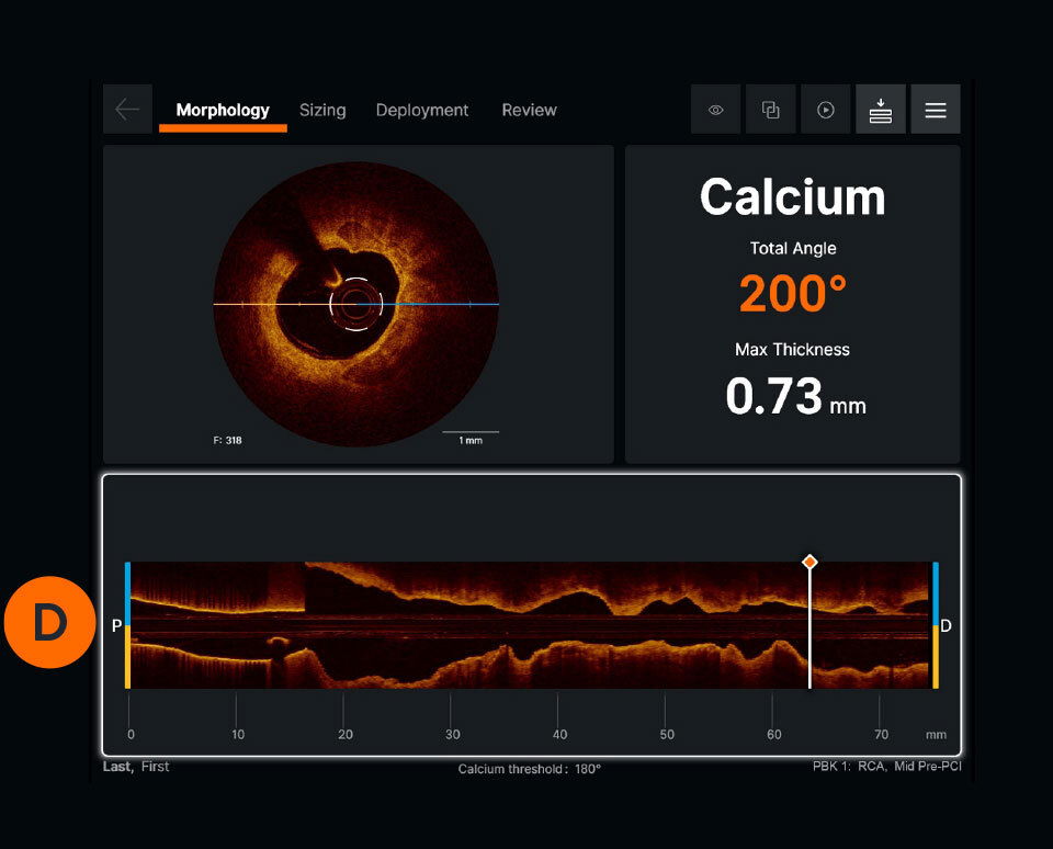
One of the main benefits of using IVUS and OCT during PCI is the ability to obtain detailed images of the coronary arteries and heart tissue. IVUS uses sound waves to create images with a resolution of about 50-100 micrometers, while OCT uses light waves to create images with a resolution of about 10 micrometers. This allows doctors to diagnose and treat heart conditions more accurately and effectively, which can improve the success rate of PCI procedures and reduce the need for repeat procedures.
In addition to providing detailed images, IVUS and OCT can also provide important information about the structure and function of the coronary arteries and heart tissue. This information is important for determining the appropriate course of treatment, such as whether PCI is necessary and which type of stent to use. For example, IVUS can help doctors identify the best location for stent placement and ensure that the stent is properly positioned, which can help prevent complications such as stent thrombosis (blood clot formation within the stent) and restenosis (re-narrowing of the artery after stent placement).
Another benefit of using IVUS and OCT during PCI is the ability to monitor the procedure in real-time. These techniques provide live images of the coronary arteries and heart tissue, which allows doctors to make immediate adjustments during the procedure if necessary. This can help prevent complications and improve the success rate of the procedure.
In addition, IVUS and OCT can provide valuable information about the health of the coronary arteries and heart tissue after the procedure has been completed. This can help doctors monitor the effectiveness of the treatment and make any necessary adjustments.
Furthermore, using IVUS and OCT during PCI can improve patient outcomes by reducing the risk of complications. These techniques provide detailed images of the coronary arteries and heart tissue, which allows doctors to identify and treat abnormalities before they become serious problems. This can help prevent complications such as stent thrombosis, restenosis, and other adverse events.
Overall, the use of IVUS and OCT during PCI offers several benefits for patients and healthcare providers. These techniques provide detailed images of the coronary arteries and heart tissue, allowing doctors to diagnose and treat heart conditions more accurately and effectively. They can also provide important information about the structure and function of the coronary arteries and heart tissue, which can help determine the appropriate course of treatment and prevent complications. In addition, the ability to monitor the procedure in real-time and monitor the health of the coronary arteries and heart tissue after the procedure can improve patient outcomes and reduce the risk of complications.